

Current in vivo imaging technology enables researchers to monitor the internal organs of laboratory animals non-invasively. This approach is considered a cutting-edge method that reduces the destructive use of experimental animals. With in vivo imaging, researchers no longer need to euthanize animals during the course of the research.
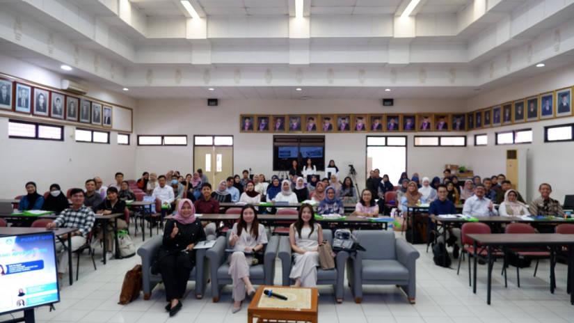
This was conveyed by veterinarian Dr. Medania Purwaningrum, Head of the Integrated Research Laboratory of the Faculty of Veterinary Medicine, Universitas Gadjah Mada (FKH UGM), during the two-day workshop titled “Veterinary Stem Cell, Cell-Free Therapy, and Modern Analytical Technology”, held on Nov. 17 to 18, 2025.
“The results can be seen directly on the device monitor. The mouse is placed inside the machine, and anyone who needs to examine its internal organs can observe them through the screen,” Dr. Purwaningrum explained at the FKH UGM Auditorium.
She noted that the increasing demand for stem cell research technologies and non-invasive analysis in veterinary medicine has encouraged the FKH UGM, through the Integrated Research Laboratory, to organize this workshop. In general, the workshop discussed various issues related to the technological needs of international-standard animal research.
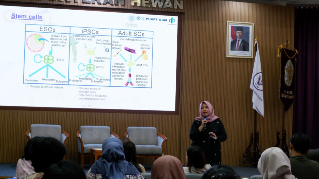
As one of the workshop speakers, Dr. Purwaningrum also introduced three key research technologies: fluorescent microscopy, the Countess cell counter, and Western blotting. The fluorescent microscope visualizes stem cells in color without the need to look through the ocular lens, as the display is already projected on a digital screen.
The Countess device is a tool for counting stem cells, allowing researchers to assess cell condition, viability, and responses to specific treatments. Meanwhile, Western blot technology is a large-scale apparatus with components valued in the billions of rupiah.
“We all hope today’s demonstration will encourage the faculty to acquire similar equipment so that our research can better align with international standards,” she remarked.
For Dr. Purwaningrum, the “Veterinary Stem Cell Workflow” workshop marks an important moment for FKH UGM in strengthening Indonesia’s modern research ecosystem. She hopes the event will not only introduce the latest technologies but also accelerate innovation in veterinary medicine and national biomedical research.
“Researchers from other faculties, such as Dentistry (FKG UGM) and Medicine, Public Health, and Nursing (FK-KMK UGM), have already begun using our facilities. Even several high schools have tried our modern research equipment. We hope this workshop expands collaboration opportunities and enhances the quality of national research,” she added.
Meanwhile, Teh Yu Xuan, a stem cell expert from Singapore, stated that protein analysis techniques have become global standards in molecular biology research. Demonstrating Western blotting techniques to workshop participants, Teh explained that the results can be obtained very quickly without the need for days of processing.
However, Teh emphasized that the characterization of animal-derived stem cells must meet the standards of the International Society for Cell & Gene Therapy (ISCT), as applied to humans.
“Everything can be completed in just four hours, and the workflow for humans and animals is essentially the same,” Teh noted.
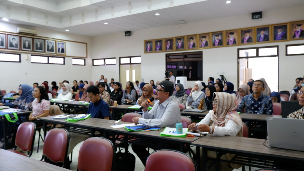
Nita, a UGM postgraduate student majoring in Veterinary Science, shared her reasons for joining the workshop. In addition to aligning with her stem cell research, she said the event provided participants with insights into emerging stem cell technologies.
“Here we learned about many new technologies that can make our laboratory work easier and more precise. That is a very valuable insight,” she said.
Author: Jelita Agustine
Editor: Agung Nugroho
Post-editor: Rajendra Arya