

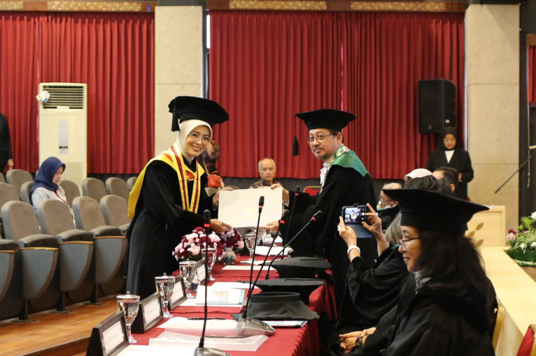
Ophthalmologist and lecturer at the Faculty of Medicine, Public Health, and Nursing Universitas Gadjah Mada (FK-KMK UGM), Reny Setyowati, has successfully developed an amniotic membrane enriched with riboflavin and moxifloxacin as an adjuvant therapy for the treatment of infectious corneal ulcers. The innovation was presented during her open doctoral examination, which investigated the effectiveness of combining the amniotic membrane with the Photoactivated Chromophore for Keratitis-Corneal Cross Linking (PACK-CXL) method to accelerate the healing of corneal infections.
Reny explained that moxifloxacin is a fourth-generation fluoroquinolone antibiotic with excellent corneal penetration. In addition, this broad-spectrum antibiotic is considered effective against both gram-positive and gram-negative pathogens.
“Moxifloxacin also has an effect on fungal corneal ulcers. Studies conducted on fungal isolates have demonstrated their ability to address fungal pathogens, particularly Fusarium,” she said during her doctoral promotion examination at FK-KMK UGM on Thursday (Jun. 11).
According to Reny, one limitation of conventional amniotic membranes is their relatively short degradation period of approximately one to two weeks. By combining the membrane with riboflavin and cross-linking therapy, the degradation period can be extended, making it more suitable for treating infectious corneal ulcers, which often require four weeks or longer to heal.
“Drug-enriched amniotic membranes can also function as a depot or slow-release system, providing the advantage of continuous and consistent antibiotic delivery for infectious corneal ulcer treatment,” she explained.

In the animal study phase, Reny employed a model of severe infectious corneal ulceration. She used rabbits inoculated with Staphylococcus aureus in accordance with clinical microbiology department standards. The results showed that the group receiving the riboflavin- and moxifloxacin-containing amniotic membrane combined with PACK-CXL experienced faster clinical improvement than the control group.
These findings were further supported by histopathological examinations. Reny reported that the intervention group demonstrated better re-epithelialization, more organized stromal structures, and fewer inflammatory cells than the control group.
“In the intervention group, the corneal epithelial layer had formed more regularly with adequate thickness, the stroma was well organized, and inflammatory cells were minimal,” she said in response to questions from her promoter.
The study also evaluated various inflammatory markers, or cytokines, in patients with infectious corneal ulcers. Reny found that changes in cytokine levels, including interleukin-6 and interleukin-1 beta, corresponded with clinical improvement in patients. These findings open opportunities for using cytokines as indicators of therapeutic success and disease progression.
According to Reny, future developments may include the use of other medications tailored to specific infectious pathogens, expanded safety testing, and applications for other corneal diseases. In the field of PACK-CXL technology, she also sees opportunities for modification, particularly in treating severe infectious corneal ulcers.
She explained that therapy could be enhanced by reducing ultraviolet irradiation time from 30 minutes to 10 minutes through protocol modifications.
In addition, future studies may explore the use of photosensitizers other than riboflavin, such as rose bengal. However, riboflavin remains the preferred option due to the substantial body of supporting research.
“We hope this will enable broader access to CXL therapy. Indonesia has a national health insurance system, so this treatment could potentially become one of the services covered under the national scheme,” she said.
Beyond presenting the research findings, Reny also outlined plans for future product development. She explained that the technology used to produce the amniotic membrane is relatively simple and can be implemented by laboratories that meet the required standards.
The research team has already secured patent certification and established collaborations with several institutions, including the National Research and Innovation Agency (BRIN) and the National Nuclear Energy Agency (BATAN), to support further development.
Dr. Setyowati’s dissertation was titled “Sediaan Membran Amnion yang Mengandung Riboflavin dan Moxifloxacin sebagai Adjuvan Terapi Photo Activated Chromophore Keratitis–Corneal Cross Linking (PACK-CXL) : Pengembangan Sediaan, Uji Preklinik dan Uji Klinik” (Riboflavin- and Moxifloxacin-Containing Amniotic Membrane Preparation as an Adjuvant to Photo-Activated Chromophore Keratitis–Corneal Cross-Linking (PACK-CXL): Formulation Development, Preclinical Testing, and Clinical Evaluation)
The open doctoral examination marked the final stage of her doctoral studies at FK-KMK UGM. She graduated with Cum Laude honors and was awarded a doctoral degree.
Her supervisory team consisted of Professor Agus Supartoto, Dr. Supanji, and Dr. Eng. Khadijah. The examination was also attended by internal and external examiners who provided input on the further development of research into the therapy of infectious corneal ulcer.
Author/Photographer: Hanifah
Editor: Gusti Grehenson
Post-Editor: Zabrina Kumara